Category: Uncategorized

  • Understanding Skin, Scars, and the Quest for Flawless Skin

    Understanding Skin, Scars, and the Quest for Flawless Skin

    Our skin is more than just a covering; itโ€™s a complex, layered organ that protects us, regulates temperature, senses the environment, and heals when injured. Yet, despite its remarkable abilities, the skinโ€™s healing process often leaves behind scars โ€” visible reminders of past wounds.


    A Quick Tour of Skin Structure

    Skin has three main layers:

    1. Epidermis โ€“ the thin, outer layer that acts as a barrier. It regenerates quickly after minor injuries.
    2. Dermis โ€“ a thicker, structural layer underneath, made of collagen and elastic fibers. This layer gives skin strength, flexibility, and volume.
    3. Subcutaneous tissue โ€“ a layer of fat and connective tissue below the dermis that cushions and supports the skin.

    The dermis is especially important when it comes to scars: loss or disruption of dermal tissue often determines whether a scar will be flat, raised, or depressed.


    How Scars Form

    When the skin is injured, the body triggers a healing cascade:

    • Clotting and inflammation stop bleeding and clear debris.
    • Fibroblasts and other cells migrate to the wound, laying down collagen and extracellular matrix.
    • The tissue gradually remodels, replacing early granulation tissue with stronger, mature collagen.

    The outcome depends heavily on dermal volume, collagen alignment, and mechanical forces in the surrounding tissue:

    • Flat scars occur when healing restores near-normal dermal thickness.
    • Raised scars (hypertrophic or keloid) result from excessive collagen deposition.
    • Depressed scars (atrophic, โ€œcraterโ€ scars) form when the dermis is lost or underfilled during healing.

    Why Some Scars Persist

    Interestingly, two injuries of similar depth may heal very differently:

    • A deep punch wound can heal perfectly flat if granulation tissue fills the defect adequately.
    • A chickenpox crater, on the other hand, often leaves a persistent depression.

    This observation highlights that dermal loss alone doesnโ€™t explain everything. Factors like inflammation, ECM destruction, local tissue tension, and the organization of collagen bundles all play a role.

    Researchers are still exploring why some scars seem to โ€œpersistโ€ even when surrounding skin looks healthy. Conventional hypotheses include:

    • Mechanical memory: the tissue around the scar may guide collagen deposition along certain lines.
    • Fibroblast positional identity: cells remember where they are in the body and what type of dermis to form.
    • Epigenetic programming: scars may leave chemical marks that influence healing.

    None of these are definitive, and many scientists emphasize that much about scar persistence remains unknown.


    Traditional Scar Treatment

    For deep or crater scars, the standard approach is surgical excision with closure:

    • The old scar is removed with a punch or scalpel.
    • The edges are brought together with sutures.
    • The new scar is often smaller, flatter, and more cosmetically favorable.

    For deeper defects, sutured closure produces cleaner, more reliable results.


    Emerging Approaches in Scar Healing

    The cutting edge of research explores ways to actively guide tissue regeneration rather than simply excising scars:

    • Electrical stimulation: Low-intensity DC or microcurrent therapy can direct cell migration (galvanotaxis), promote fibroblast activity, and enhance angiogenesis. By guiding cells into a wound, it may improve dermal filling and flatten depressed scars.
    • Mechanical remodeling: Techniques like subcision or tension-controlled closure influence collagen deposition and scar shape.
    • Tissue scaffolds and growth factors: These can provide a framework for cells to rebuild dermal volume more effectively.

    While many of these strategies are still in research or clinical trials, they hint at a future where scars may not just be minimized, but actively regenerated into normal-looking skin.


    The Takeaway

    Skin is remarkable, but healing is a complex balancing act that we currently don’t fully understand. Whether a scar is flat, raised, or depressed depends on a combination of tissue volume, cellular behavior, collagen organization, and mechanical forces.

    Modern science is now uncovering ways to guide these processes, from careful surgical techniques to electrical stimulation and regenerative scaffolds. Though we donโ€™t fully understand every mechanism, the future of scar healing looks increasingly active, precise, and regenerative.


  • The Science of Titanium Dental Implants

    The Science of Titanium Dental Implants

    1. What makes dental implants biocompatible?

    2. What can improve biocompatibility?

    3. What are the potential downsides?

    Our bodies do not reject titanium. The discovery is attributed to Perโ€Ingvar Branemark, a Swedish physician who implanted a titanium screw into a bunny bone and reported no rejection by the surrounding tissue (1). To use Hollywood jargon in regards to animals in movies: โ€œThe bunny was seriously hurt in the process of โ€˜filmingโ€™ the 1969 paperโ€.

    How did he come up with the idea to use a titanium screw, one may rightfully ask? This is because there was some previous knowledge about titaniumโ€™s properties published before World War II. The researchers implanted various metals into lab animalsโ€™ bones and found that titanium was โ€œspecialโ€ in a good way (2). The discovery is not attributed to Bothe, Beaton, and Davenport, who published this, but rather to the person who did more or less the same thing 29 years later.

    Branemark didnโ€™t get recognition right after his discovery. He tried to secure funding for his research but couldnโ€™t win grants from his own people. The scientific community couldnโ€™t accept that a metal could be biocompatible. Luckily, NIH gave him a grant to continue his research, and finally, in 1982, he demonstrated to the American Dentist Association that titanium teeth implants work (3). His research didnโ€™t result in a Nobel Prize, regardless of the fact that he was of the right gender and the right ancestry for the time period. His discovery apparently didnโ€™t seem original enough to the Nobel Committee. Since he was at the epicenter of action (Sweden), one canโ€™t help but speculate that the decision-makers knew him and didnโ€™t like something about him, which is always a factor when impartial decisions are made. Well, he did file for many patents in the early days of implant surgery. Not bad, really. If something was hurt, it was probably just the ego.

    Titanium is not inert at all. It reacts with the surrounding tissues, but it seems that the body doesnโ€™t mind it. The important feature of this metal is its ability to coat its surface with a thin layer of oxide, thus preventing corrosion. When titanium is exposed to body fluids, the oxide on the surface becomes hydrated, so the surface is covered with Ti-OHmolecules that are negatively charged, attracting Ca2+ ions that lead to the formation of apatite layer as there are phosphate ions around as well. Hydroxyapatite is a natural component of bones; hence, it is biocompatible. This is a somewhat simplified explanation, but the basic mechanism is correct. We believe in not overwhelming the readers with details right at the beginning because once the underlying mechanism is understood, it is easy to fill in the details later.

    The process is not as simple as if a smooth, shiny metal were drilled into the bone. In fact, the surface of the titanium screw is heavily modified. Take a look at the image. The screw part of the implant is titanium, but it looks white and rough because of the oxide layer. To achieve this effect, implant manufacturers use different approaches. There are two most common metal treatments: SLA and PSC.

    SLA stands for โ€œSand blast Larger grit and Acid etchingโ€ (4). The treatment starts with large abrasive particles (5-20 microns), then acid treatment, after which another sandblasting with a particle range of 3-5 microns is applied, followed by another treatment with sulfuric acid. The specifics of the protocol were probably developed based on empirical evidence, as electron microscopy showed that osteoblasts prefer to colonize a uniformly etched surface as opposed to an irregular one. This uniformity is achieved with an acid treatment. Sandblasting alone produces irregular surface and it’s not enough.

    Another very common surface treatment is Plasma Spray Coating. Materials such as hydrohyapatite are heated to high temperatures (9000 ยฐC) and deposited on the surface of the implant. Titanium can also be deposited on the surface as well. Since the goal is to achieve adequate roughness and uniformity, the initially strange-sounding idea of depositing titanium on titanium eventually makes sense. Plasma coating is used more frequently than the SLA method.

    The previous two methods started with a smooth metal surface. There is an alternative approach. One can produce a porous titanium implant because it can improve osseointegration (colonization by osteoblasts). The downside is a messy surgery if it ever needs to be removed.

    The efficiency of the surface treatments is evaluated with Simulated Body Fluids solution. As the name suggests, this solution mimics the environment of the body. It was shown that plasma-coated hydrohyapatite is less durable and makes less contact with the tissue than titanium coating (4,5). This is strange because it is the natural bone material, so could it be that the result was observed due to the technique itself, not the intrinsic quality of the material used? Our bones are not titanium-strong. Hydrohyapatite should be strong enough and more readily accepted than the foreign metal. Strange results…

    After the surface of an implant is treated with SLA or PSC, the implant is soaked in sodium hydroxide. This step enhances the production of amorphous sodium titanate in the form of a hydrogel. Once heated, it dries, becoming more dense, which is desirable. The surface will be more hydrophilic once it is back in the solution. Surface โ€œwettingโ€ is essential for the integration of an implant into the surrounding tissue. Material that is hydrophobic or inert will not get coated with body fluids and will not attract osteoblasts for colonization.

    So far, we have discussed only the chemistry of the process, not biology or timing. Within 24 hours of the surgery, the implant site is populated only by red blood cells and macrophages (6). After 96 hours, the site is visited by mesechymal stem cells. New blood vessels are being formed. After four weeks, osteoblasts colonize the area and finally bridge the implant with the surrounding tissue after approximately six to eight weeks.

    Zirconium belongs to the same group as titanium in the periodic table. Their properties should be similar. Still, metal zirconium is not used for manufacturing implants but rather zirconium dioxide or zirconia. Being a white ceramic material, it has the advantage of appearance, but titanium shows better mechanical properties (7). In terms of biocompatibility, they are comparable, provided that proper surface treatment has been applied. With no prior surface treatment, titanium is superior, but that might be a moot point.

    There seem to be no additional candidates that can replace titanium or zirconia for now. For that reason, improvements to surface treatment methods will most likely be a major thing in the near future. One thing that is important but has not been discussed so far is bacterial infection. If the implant does not adhere well to the surrounding tissues, bacteria will, and once they form a biofilm, they can cause severe infections, leading to the loss of the implant. Preventing bacterial infections is one direction that may improve the longevity of implants. An elegant method has been devised. It consists of doping titanium with copper (8). Small amounts of copper are not toxic to us, but localized release at the implant site prevents bacterial growth. Neat!

    This world would not be as it is if there were no data in direct opposition to what we have said so far. New research shows that both titanium and titanium dioxide might be toxic. The first thing that comes to mind is silica. If you need pure silica for the lab, it will come in a container with a label saying โ€œcarcinogenโ€. People who do metal casting use fine silica, and they are in real danger of developing silicosis and lung cancer (9). Still, we donโ€™t wear a mask once we go to the beach. This statement has been challenged lately by some Darwin Award candidates.

    We have had decades worth of data that shows one thing, and now we have some new, potentially deeper insights into why the previous observations were not perfect. The potential harmful effects of TiO2 are more related to the large amounts of nanoparticles that we use as a food additive and sunscreen (10). The amounts of titanium dioxide on the surface of an implant are tiny in comparison to what is used in food or cosmetics, but the negative effects do come from the metal itself.

    One downside of titanium implants is hypersensitivity in some patients (11). A patch test used to determine allergy sounds logical, but it doesnโ€™t correlate with the clinical outcome (12).

    In addition to allergies, titanium can cause the nails to turn yellow – the yellow nail syndrome (13). There have been reports of the accumulation of titanium in organs and chronic sinusitis. The main toxicity- and allergy-producing components are the metals used to make the titanium alloy, such as aluminum and vanadium. These metals are common in modern titanium implants. For an in-depth review of titanium toxicity, we recommend the following reference (12). Someone who has no knowledge of metallurgy would now ask, โ€œBut why donโ€™t they use pure titanium?โ€. No, it is not practical. Working with pure metals (other than gold) is a pain unlike many others. Pure metals crystallize and have a very sharp melting point that is usually higher than when alloyed. Machining is more difficult because pure metals tend to be brittle, so alloys are always better. In this particular case, the metals added to titanium should be tissue-friendly. Most are not.

    Manufacturers claim that the implant can last 25 years or even longer and that the failure is due to bone loss because implants are usually used in advanced-age patients. It would be interesting to see a study with individuals who had dental implants much earlier in life.

    1. Branemark PI, Adell R, Breine U, Hansson BO, Lindstrom J, Ohlsson A. Intra-osseous anchorage of dental prostheses. I. Experimental studies. Scand J Plast Reconstr Surg. 1969;3(2):81-100. doi: 10.3109/02844316909036699. PMID: 4924041.

    2. Jokstad A. Why did Professor Per-Ingvar Brรฅnemark never receive the Nobel Prize in Medicine? Clin Exp Dent Res. 2017 Jul 3;3(3):79-80. doi: 10.1002/cre2.72. PMID: 29744182; PMCID: PMC5719826.

    3. Kim TI. A tribute to Dr. Per-Ingvar Brรฅnemark. J Periodontal Implant Sci. 2014 Dec;44(6):265. doi: 10.5051/jpis.2014.44.6.265. Epub 2014 Dec 31. PMID: 25568805; PMCID: PMC4284373.

    4. Jemat A, Ghazali MJ, Razali M, Otsuka Y. Surface Modifications and Their Effects on Titanium Dental Implants. Biomed Res Int. 2015;2015:791725. doi: 10.1155/2015/791725. Epub 2015 Sep 7. PMID: 26436097; PMCID: PMC4575991.

    5. Yang GL, He FM, Yang XF, Wang XX, Zhao SF. Bone responses to titanium implants surface-roughened by sandblasted and double etched treatments in a rabbit model. Oral Surg Oral Med Oral Pathol Oral Radiol Endod. 2008 Oct;106(4):516-24. doi: 10.1016/j.tripleo.2008.03.017. Epub 2008 Jul 7. PMID: 18602288.

    6. Silva RCS, Agrelli A, Andrade AN, Mendes-Marques CL, Arruda IRS, Santos LRL, Vasconcelos NF, Machado G. Titanium Dental Implants: An Overview of Applied Nanobiotechnology to Improve Biocompatibility and Prevent Infections. Materials (Basel). 2022 Apr 27;15(9):3150. doi: 10.3390/ma15093150. PMID: 35591484; PMCID: PMC9104688.

    7. Hanawa T. Zirconia versus titanium in dentistry: A review. Dent Mater J. 2020 Jan 31;39(1):24-36. doi: 10.4012/dmj.2019-172. Epub 2019 Oct 30. PMID: 31666488.

    8. Wu Y, Zhou H, Zeng Y, Xie H, Ma D, Wang Z, Liang H. Recent Advances in Copper-Doped Titanium Implants. Materials (Basel). 2022 Mar 22;15(7):2342. doi: 10.3390/ma15072342. PMID: 35407675; PMCID: PMC8999642.

    9. Sato T, Shimosato T, Klinman DM. Silicosis and lung cancer: current perspectives. Lung Cancer (Auckl). 2018 Oct 26;9:91-101. doi: 10.2147/LCTT.S156376. PMID: 30498384; PMCID: PMC6207090.

    10. Racovita AD. Titanium Dioxide: Structure, Impact, and Toxicity. Int J Environ Res Public Health. 2022 May 6;19(9):5681. doi: 10.3390/ijerph19095681. PMID: 35565075; PMCID: PMC9104107.

    11.Poli PP, de Miranda FV, Polo TOB, Santiago Jรบnior JF, Lima Neto TJ, Rios BR, Assunรงรฃo WG, Ervolino E, Maiorana C, Faverani LP. Titanium Allergy Caused by Dental Implants: A Systematic Literature Review and Case Report. Materials (Basel). 2021 Sep 12;14(18):5239. doi: 10.3390/ma14185239. PMID: 34576463; PMCID: PMC8465040.

    12. Kim KT, Eo MY, Nguyen TTH, Kim SM. General review of titanium toxicity. Int J Implant Dent. 2019 Mar 11;5(1):10. doi: 10.1186/s40729-019-0162-x. PMID: 30854575; PMCID: PMC6409289.

    13. Decker A, Daly D, Scher RK. Role of Titanium in the Development of Yellow Nail Syndrome. Skin Appendage Disord. 2015 Mar;1(1):28-30. doi: 10.1159/000375171. Epub 2015 Feb 11. PMID: 27172293; PMCID: PMC4857837.

  • Aflatoxins – What We Know So Far

    Aflatoxins – What We Know So Far

    What are Aflatoxins?

    Aflatoxins are metabolite products of Aspergillus, a genus of fungi frequently present in grain foods such as wheat, corn, rice, as well as peppers, ground nuts, tree nuts, and hay. Hay, of course, is not our food, but it can affect us indirectly.

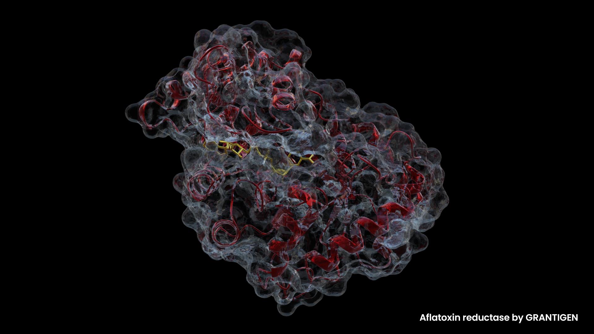
    There are over 400 micotoxins, of which Aflatoxin B1 (AFB1), one of the 21 aflatoxins, is the most harmful (1).

    It is also the most important due to its deleterious effects on both our health and the food industry’s profit. Unfortunately, aflatoxins are thermally stable. Cooking does not degrade them (2).

    ABF1 causes hepatotoxicity, nephrotoxicity, neurotoxicity, immunosuppression and adversely affects reproduction in laboratory animals. Most people have heard of aflatoxin in milk. The milk aflatoxin is designated as AFM1, and it is the hydroxylated version of AFB1 that is produced by dairy cows who eat contaminated feed (3). Both are classified as carcinogens, with M1 being slightly less toxic.

    What is Aflatoxins’ mechanism of action?

    The mechanism of action is complicated because the toxin affects many cellular pathways. As AFB1 gets processed in the liver, it gets oxidized by the P450 family of enzymes, forming a reactive epoxide. The epoxide readily binds DNA causing mutations (4).

    Nothing that changes DNA is good for health. However, we were all taught that random mutations in DNA lead to gain of function and ultimately transition from species to species. How else would one explain the evolution of humans from monkeys unless, at some point in distant history, a random mutation produced an โ€œugly monkeyโ€ with a higher brain capacity, less fur, or the ability to walk upright, talk, or all of the above? That weird monkey must have happened by chance-say, one in a million. Itโ€™s safe to assume that human’ ancestors didn’t care about diversity so the poor monkey was probably ridiculed and unable to find a mate among the regular monkeys. Luckily, it must have found another โ€œone in a million monkeyโ€ of the opposite sex within walking distance and produced offspring that built the foundation for our species.

    We donโ€™t know what triggered this serendipitous transformation in monkey DNA, especially what caused two pairs of their chromosomes to merge into one, as we have one pair of chromosomes less. Guessing that aflatoxins played a role in this is a guess as good as any other. These compounds are potent DNA modification agents, and DNA change is needed for the transformation of species. Maybe aflatoxins are not that bad after all? Maybe we owe them our existence? Shakespeare wrote, โ€œThere is nothing either good or bad, but thinking makes it so.โ€ This perfectly aligns with evolution. Evolution knows neither good nor bad; it just happens. Therefore, we may not like dying of cancer, but itโ€™s not relevant. Isnโ€™t it interesting how painful it can be to observe oneโ€™s world from a non-anthropocentric point of view?

    What is relevant, though, is that random mutations happen, and while most of the time they cause death and disability, occasionally they produce something new and something good. At least there is scientific consensus on that, regardless of the fact that we canโ€™t see it happening in real life. Crooked-beaked finches and dark moths donโ€™t really count, as those examples are weak.

    After this detour into the possible mechanism of monkey-to-human transformation based on solid science doped with aflatoxins’ speculative role in evolution, we can now continue listing additional ways in which aflatoxins can harm us.

    The toxin also depletes glutathione. Since glutathione is responsible for detoxifying the body, AFB1 can bind glutathione, causing elevated levels of reactive oxygen species due to the lack of this important endogenously produced antioxidant. For those who have wondered why itโ€™s dangerous to take more Tylenol than what is written on the drug leaflet, the answer is the same. Tylenol too sequesters the slow-producing glutathione, leading to oxidative stress in mitochondria and ultimately triggering apoptosis (5).

    Together with these two potential primary means of action, ABF1 affects steroid hormone biosynthesis, rethionoid and lipid metabolism, as well as AMPK, cAMP, insulin, mTOR, VEGF, FoxO, MAPK, TNF signaling pathways, just to name a few. The outcomes of large aflatoxin consumption could be liver cancer, lung cancer, and gastrointestinal cancer. The full list of pathways affected by aflatoxins is much longer. Readers can find out more from this review (6).

    How is it possible that one molecule or one set of closely related molecules can affect so many biological processes? It seems that ABF1 is a Swiss army knife, but in a sinister way. It must be because it looks like something normally found in metabolism. Obviously, it shares an (evolutionary) design with steroid hormones and vitamin D. This could explain its interference with so many pathways. The full explanation will hopefully come in the near future because, although the author of this post cares about future generations, he cares a bit more about his health right now.

    Another possible structure/function similarity interplay might be inferred from the fact that AFB1 downregulates the expression of the vitamin D receptor in the human osteasacroma cell line SAOS-2. Maybe that is why rickets is possible in places like Africa with abundant sunshine throughout the year, assuming an adequate diet is available (7)?

    Let us now focus on the practical problems, such as:

    1. Is it possible to remove aflatoxins from food?

    2. How can we find out whether we have accumulated aflatoxins in our bodies?

    3. Is it possible to detoxify from them?

    1. Is it possible to remove aflatoxins from food?

    It is, but with various degrees of success depending on the method (8). The first method is absorption on various types of materials such as charcoal. It is a physical method of low efficiency. In order to improve adsorption, the current adsorbents need to be improved. There have been continued efforts in this direction.

    The second method is the chemical treatment of food. Itโ€™s more aggressive and efficient, but treating food with acids, bases, and oxidative agents changes the food as well, not just the toxin. Therefore, this method is not ideal either. Sodium metabisulphite and hydrogen peroxide combination seems to work quite well (9).

    The third method relies on โ€œgoodโ€ microorganisms that can perform the detoxification of food (10). Good microorganisms can also absorb aflatoxins and degrade them. Food production processes may benefit from using good microorganisms, but at the same time, it may not be feasible to grow them on food just to prevent potential contamination with Aspergillus. Still, the microbial method is better than the previous two.

    2. How can we find out whether we have accumulated aflatoxins in our bodies?

    It is possible to detect accumulated toxins through the detection of their metabolites in urine. As we have already mentioned, AFM1 is excreted in milk, so it can be analyzed there. Blood can be analyzed as well, with various degrees of success due to the sensitivity of the method used (11).

    3. Is it possible to detoxify our bodies?

    Apparently yes, but the protocol has not yet been established. Curcumin has emerged as a potential scavenger, or better said, a protective agent for AFB1 (12). Curcumin has been investigated for many positive effects on health through its antibacterial, antiviral, and antitumor properties. The health benefits of this molecule go beyond what is listed here.

    So far, curcuminโ€™s protective actions have been investigated in vitro using BHF12 and HUC-PC cell lines, mice, rats, tilapia fish, chickens, and ducklings of different ages. In each case, it was determined that the group of animals ingesting curcumin in their diet contaminated with AFB1 had less tissue damage than the control group. Readers can find out more details in (12).

    Grantigen needs to note that the experimental conditions used in the research mentioned do not closely mimic a real-life scenario because the amounts of both curcumin and AFB1 used were much larger than what would be the case in a real-life scenario. Even the ratio of the two may not necessarily be proportionately higher than what could happen in a regular diet in order to say that the experimental design just augmented the real-life scenario.

    For example, in one study, ducklings were fed 400 mg/kg of curcumin per day. For a human of 70kg, that would be 28g of curcumin per day. Since it doesnโ€™t come in pure form but rather in turmeric powder that contains about 3% of the active component, the amount of turmeric powder would end up being 933 g per day! This is the high end of the experiment design range. According to the duckling massacre paper, the protective effects of curcumin were confirmed in the tissues of 21-day-old ducklings (12). Apart from its macabre nature, this is a really good review that we recommend to anyone who wants to learn more on the topic but doesnโ€™t have time to sift through the mountain of literature on aflatoxins.

    In some other experiments, chickens were fed with the amount of turmeric that would be equivalent to 31 g of turmeric per day, which for an average human is possible. Considering the other beneficial effects of curcumin, this amount of turmeric ingested per day could potentially be a good addition to the western diet. Can it go much lower and still work?

    Despite all of this, the best way to protect oneself from aflatoxins is to avoid them. It is more or less possible, but it also greatly depends on the region of the world. For example, hot and humid places are more likely to have elevated amounts of toxins in food, especially if this is coupled with lax food regulations in that country. In contrast, cold and arid countries that are regulated โ€œto the bone,โ€ like northern Europe and Canada, are less likely to have the toxin lurking in food, as that is one of the things that is checked before food is brought to consumers. Milk is easy to avoid because everyone who is not an infant can easily live without it. Yogurt contains the good microbes that are Aspergillusโ€™ natural enemies; therefore, it could be safer than milk. Grains are everywhere, so they are difficult to avoid, and for that reason, one would end up relying on the government to protect him, which is always a scary thought.

    Let us finish on a positive note:

    So what happens if we consume low amounts of aflatoxin over a long period of time? The amounts that are not going to produce acute poisoning According to the study on 131 patients conducted in Italy, where in certain regions contamination is known to be present, but at low levels, 81% of hepatocellular carcinoma patients did not have traces of AFB1 in the liver cells (13). Of the remaining 19% who had traces of AFB1, only a few had high levels of the toxin in the cells. This study does not support a link between hepatocellular carcinoma and low ABF1 levels. Where was the negative control in this study anyway? Wouldnโ€™t a negative control composed of healthy people who could potentially have mixed results in regards to traces of ABF1 in liver cells completely destroy the fear of low doses of aflatoxins? This may not be a good study to publish for a lab that gets funded to study aflatoxins, hence the lack of negative control.

    Another study found aflatoxin present in the sera of 64% of primary liver cancer patients, while at the same time finding hepatitis B in 69% of the patients (14). This shows a strong correlation between the two, but the study doesnโ€™t tell us what the correlation would be if there was no hepatitis B present. The study was conducted in Sub-Saharan Africa, a region that is at high risk of food contamination by Aspergillus; therefore, the comparison with the European study is not easy, or maybe not possible at all. We may have missed the paper we were looking for, but to Grantigen, it seems that the definitive link between low doses of aflatoxin exposure and hepatocellular carcinoma in the absence of other risk factors has not been established.

    References:

    1. Bennett JW, Klich M. Mycotoxins. Clin Microbiol Rev. 2003 Jul;16(3):497-516. doi: 10.1128/CMR.16.3.497-516.2003. PMID: 12857779; PMCID: PMC164220.

    2. Yamada M, Hatsuta K, Niikawa M, Imaishi H. Detoxification of Aflatoxin B1 Contaminated Maize Using Human CYP3A4. J Microbiol Biotechnol. 2020 Aug 28;30(8):1207-1213. doi: 10.4014/jmb.2003.03032. PMID: 32423188; PMCID: PMC9728267.

    3. Dhakal A, Hashmi MF, Sbar E. Aflatoxin Toxicity. 2023 Feb 19. In: StatPearls [Internet]. Treasure Island (FL): StatPearls Publishing; 2023 Janโ€“. PMID: 32491713.

    4. Johnson WW, Guengerich FP. Reaction of aflatoxin B1 exo-8,9-epoxide with DNA: kinetic analysis of covalent binding and DNA-induced hydrolysis. Proc Natl Acad Sci U S A. 1997 Jun 10;94(12):6121-5. doi: 10.1073/pnas.94.12.6121. PMID: 9177180; PMCID: PMC21012.

    5. Benkerroum N. Chronic and Acute Toxicities of Aflatoxins: Mechanisms of Action. Int J Environ Res Public Health. 2020 Jan 8;17(2):423. doi: 10.3390/ijerph17020423. PMID: 31936320; PMCID: PMC7013914.

    6. Marchese S, Polo A, Ariano A, Velotto S, Costantini S, Severino L. Aflatoxin B1 and M1: Biological Properties and Their Involvement in Cancer Development. Toxins (Basel). 2018 May 24;10(6):214. doi: 10.3390/toxins10060214. PMID: 29794965; PMCID: PMC6024316.

    7.Costanzo P, Santini A, Fattore L, Novellino E, Ritieni A. Toxicity of aflatoxin B1 towards the vitamin D receptor (VDR). Food Chem Toxicol. 2015 Feb;76:77-9. doi: 10.1016/j.fct.2014.11.025. Epub 2014 Dec 4. PMID: 25483621.

    8. Guan Y, Chen J, Nepovimova E, Long M, Wu W, Kuca K. Aflatoxin Detoxification Using Microorganisms and Enzymes. Toxins (Basel). 2021 Jan 9;13(1):46. doi: 10.3390/toxins13010046. PMID: 33435382; PMCID: PMC7827145.

    9. Karlovsky P, Suman M, Berthiller F, De Meester J, Eisenbrand G, Perrin I, Oswald IP, Speijers G, Chiodini A, Recker T, Dussort P. Impact of food processing and detoxification treatments on mycotoxin contamination. Mycotoxin Res. 2016 Nov;32(4):179-205. doi: 10.1007/s12550-016-0257-7. Epub 2016 Aug 23. PMID: 27554261; PMCID: PMC5063913.

    10. Yamada M, Hatsuta K, Niikawa M, Imaishi H. Detoxification of Aflatoxin B1 Contaminated Maize Using Human CYP3A4. J Microbiol Biotechnol. 2020 Aug 28;30(8):1207-1213. doi: 10.4014/jmb.2003.03032. PMID: 32423188; PMCID: PMC9728267.

    11. Wild CP, Chapot B, Scherer E, Den Engelse L, Montesano R. Application of antibody methods to the detection of aflatoxin in human body fluids. IARC Sci Publ. 1988;(89):67-74. PMID: 3198233.

    12. Dai C, Tian E, Hao Z, Tang S, Wang Z, Sharma G, Jiang H, Shen J. Aflatoxin B1 Toxicity and Protective Effects of Curcumin: Molecular Mechanisms and Clinical Implications. Antioxidants (Basel). 2022 Oct 14;11(10):2031. doi: 10.3390/antiox11102031. PMID: 36290754; PMCID: PMC9598162.

    13. Gramantieri L, Gnudi F, Vasuri F, Mandrioli D, Fornari F, Tovoli F, Suzzi F, Vornoli A, D’Errico A, Piscaglia F, Giovannini C. Aflatoxin B1 DNA-Adducts in Hepatocellular Carcinoma from a Low Exposure Area. Nutrients. 2022 Apr 15;14(8):1652. doi: 10.3390/nu14081652. PMID: 35458213; PMCID: PMC9024438.

    14. Tchana AN, Moundipa PF, Tchouanguep FM. Aflatoxin contamination in food and body fluids in relation to malnutrition and cancer status in Cameroon. Int J Environ Res Public Health. 2010 Jan;7(1):178-88. doi: 10.3390/ijerph7010178. Epub 2010 Jan 18. PMID: 20195440; PMCID: PMC2819783. (63% in liver cancer)

  • Can Metformin extend life?

    Can Metformin extend life?

    In order not to waste paragraphs and readersโ€™ time, let us answer the question: If you have type 2 diabetes, metformin will indeed increase your lifespan because it treats the condition that shortens it. If you are healthy, and especially if you workout, metformin will not increase your lifespan. It is not a miracle drug because it was proven that diet and exercise have stronger positive effects on health. The beneficial effects do not add up. In fact, metformin makes exercise less effective (1) . There, you donโ€™t need to read more. We just summarized the studies addressing this question for you. The exact pathways are not really important if you are not interested in the molecular mechanisms of our bodies.

    However, new data always comes up, and who knows what the future holds? Maybe metformin in combination with, say, cinnamon will improve the already improved bodies through diet and exercise? This is the beauty of not knowing everything. One just needs to be open to new ideas and not heavily invested in the current state of things, as that always makes one reluctant to accept anything new.

    Here is a more detailed explanation:

    The second section of the article is for those who want a leisurely read with enough scientific information along the way.

    Metformin was first synthesized in 1922 because there was some previous knowledge that the โ€œtoxic weedโ€ Galega officinalis may help with diabetes (2). Diabetes was not understood in the ye olde times, but people did know that some disease that causes sweet urine existed. How did one find out whether one fellowโ€™s urine was sweet? How did people find out that a particular plant among so many others could help with this particular condition?

    It took some time before metformin was approved and prescribed for type 2 diabetes. It has been 70 years since then. The drug is considered safe and effective, but the interesting thing is that the exact mechanism of action is not fully understood. There have been many articles that are trying to pinpoint the exact mechanism, though it is known which cellular pathways it affects (3).

    This would be a good time to say that metformin is off-patent now and also very cheap. There are efforts to create something new, patented, and way more expensive that will do the same job with possibly more side effects. The side effects can be easily mitigated with an appropriate advertising campaign. Humans are so predictable…

    Our understanding of biological processes on the molecular level keeps growing. With that, the number of possible signaling pathways that need to be investigated also grows. Researchers have tried to figure out the role of metformin in the AMP-activated protein kinase (AMPK) signaling pathway, mTor pathway, IGF1 pathway, GLP1 pathway, DAF16 pathway, CBP pathway, and the role it may play in inhibiting mitochondrial complex I.

    We can briefly summarize the findings here, but readers can find out more from (4).

    1. The AMPK pathway is named after the 5′ adenosine monophosphate-activated protein kinase. As the name suggests, it is a kinase activated by adenosine monophosphate. Its function is to serve as a molecular energy sensor in cells. AMPK senses energy by comparing the ratio of adenosine monophosphate (AMP) to adenosine triphosphate (ATP). When the AMP/ATP ratio increases, it sets in motion processes of ATP production and glucose uptake. AMPK also regulates lipid metabolism processes. Metformin activates AMPK, which has positive effects on cell function, but as we mentioned before, so does exercise. (5)

    2. The mammalian target of rapamycin (mTOR) plays an important role in proliferation and immune cell differentiation. It is activated in tumors. Metformin inhibits this pathway through the AMPK pathway, resulting in a lesser incidence of cancer, as diabetes is a risk factor for cancer (6).

    3. Insulin-like growth factors (IGFs) affect cell proliferation, differentiation, and survival. Although this signaling pathway is considered โ€œweakโ€ by itself (7) and primarily augments other pathwaysโ€™ signals, it too is affected by metformin (negatively), thus mimicking the calorie restriction situation that has been proven to increase lifespan. What would be the alternative to taking metformin in this particular case? Eating less, perhaps?

    4. Glucagon-like peptide 1 (GLP1) has a role in the regulation of glucose levels by insulin secretion. Metformin enhances the release of GLP1, thus reducing glucose levels. (8).

    5. DAF16 is specific for C. elegans, as many longevity experiments have been done on this nematode. The gene is involved in glucose and lipid metabolism. Metformin increases insulin sensitivity, which simulates a reduced calorie intake. Too much sugar in the diet leads to insulin insensitivity, and vice versa. C. elegans has a very different gut lining than humans, making drug uptake less efficient. For that reason, metformin concentrations in experiments with C. elegans are up to a thousand times higher than in humans. There is an up to 36% increase in lifespan in the worm that was attributed to the positive effect methformin has on its gut flora, i.e., killing the wormโ€™s bacteria. The antibiotic trimetoprim had the same effect on the worms (3). Should we all take Bactrim to increase our lifespan?

    6. AMPK-atypical protein kinase C (aPKC)-CREB-binding protein (CBP) has neuroprotective properties and a role in cell proliferation, growth, and apoptosis. CREB-binding protein (CBP), as its name suggests, binds CREB (a transcriptional factor), making it active. Metformin and insulin cause the dissociation of CBP from the promoter region, which results in halted expression of the downstream genes responsible for gluconeogenesis. In other words, metformin stops the synthesis of glucose through this pathway (9).

    7. Mitochondrial complex I is the first complex in the respiratory chain of mitochondria. When metformin inhibits complex I, it leads to a higher AMP/ATP ratio (see above) and activates the AMPK pathway (4). However, these results were obtained in cell cultures. When tested in mice, mitochondrial activity was elevated. There is some kind of interaction between metformin and complex I, but it would be strange to learn that decreasing respiration leads to a positive outcome for the cell. It is well known that young cells have a higher respiration rate than old ones (10).

    Metformin is a great example of how something from nature was translated into an effective drug. The only other class of drugs that come to mind are antibiotics. With so many articles written about this drug, it is normal to have high expectations, but doesnโ€™t it already treat the condition for which it was intended? People just want more…

    Science is close, but not always in a clear-cut scenario. We are constantly in the process of finding out new information. There have been examples when a solid scientific truth was shaken by new data and then eventually destroyed. This happens very slowly because of the opposition, which has vested interests, of course. The landscape of the world where useful information lives is cluttered with opposing statements and interests that constantly create noise. It takes a sharp mind with enough scientific background to cut through the noise and get to the truth. Some people are lucky to be born with a natural sense for that thing, usually called common sense. Others must go through the process of practicing the skill. The third group never masters it. Not to deviate too much from the topic of the post, we should say that metformin is a great drug that unfortunately doesnโ€™t fulfill the hopes of extending life. Not changing oneโ€™s lifestyle and only relying on a single molecule to live longer is tempting for many people. Metforminโ€™s role in cancer prevention has not been researched as thoroughly. Maybe it possesses some serious anti-cancer properties that we are not yet aware of?

    References:

    1.Walton RG, Dungan CM, Long DE, Tuggle SC, Kosmac K, Peck BD, Bush HM, Villasante Tezanos AG, McGwin G, Windham ST, Ovalle F, Bamman MM, Kern PA, Peterson CA. Metformin blunts muscle hypertrophy in response to progressive resistance exercise training in older adults: A randomized, double-blind, placebo-controlled, multicenter trial: The MASTERS trial. Aging Cell. 2019 Dec;18(6):e13039. doi: 10.1111/acel.13039. Epub 2019 Sep 26. Erratum in: Aging Cell. 2020 Mar;19(3):e13098. PMID: 31557380; PMCID: PMC6826125.

    2. Bailey CJ, Day C. Metformin: its botanical background. Pract Diab Int. 2004 Apr 21;3

    3. Glossmann HH, Lutz OMD. Metformin and Aging: A Review. Gerontology. 2019;65(6):581-590. doi: 10.1159/000502257. Epub 2019 Sep 13. PMID: 31522175.

    4. Mohammed I, Hollenberg MD, Ding H, Triggle CR. A Critical Review of the Evidence That Metformin Is a Putative Anti-Aging Drug That Enhances Healthspan and Extends Lifespan. Front Endocrinol (Lausanne). 2021 Aug 5;12:718942. doi: 10.3389/fendo.2021.718942. PMID: 34421827; PMCID: PMC8374068.

    5. Richter EA, Ruderman NB. AMPK and the biochemistry of exercise: implications for human health and disease. Biochem J. 2009 Mar 1;418(2):261-75. doi: 10.1042/BJ20082055. PMID: 19196246; PMCID: PMC2779044.

    6.Shahid RK, Ahmed S, Le D, Yadav S. Diabetes and Cancer: Risk, Challenges, Management and Outcomes. Cancers (Basel). 2021 Nov 16;13(22):5735. doi: 10.3390/cancers13225735. PMID: 34830886; PMCID: PMC8616213.

    7. Hakuno F, Takahashi SI. IGF1 receptor signaling pathways. J Mol Endocrinol. 2018 Jul;61(1):T69-T86. doi: 10.1530/JME-17-0311. Epub 2018 Mar 13. PMID: 29535161.

    8. Drucker DJ. Mechanisms of Action and Therapeutic Application of Glucagon-like Peptide-1. Cell Metab. 2018 Apr 3;27(4):740-756. doi: 10.1016/j.cmet.2018.03.001. PMID: 29617641.

    9. Gouveia A, Hsu K, Niibori Y, Seegobin M, Cancino GI, He L, Wondisford FE, Bennett S, Lagace D, Frankland PW, Wang J. The aPKC-CBP Pathway Regulates Adult Hippocampal Neurogenesis in an Age-Dependent Manner. Stem Cell Reports. 2016 Oct 11;7(4):719-734. doi: 10.1016/j.stemcr.2016.08.007. Epub 2016 Sep 8. PMID: 27618724; PMCID: PMC5063627.

    10. Sun N, Youle RJ, Finkel T. The Mitochondrial Basis of Aging. Mol Cell. 2016 Mar 3;61(5):654-666. doi: 10.1016/j.molcel.2016.01.028. PMID: 26942670; PMCID: PMC4779179.

    11. Zeng YC, Sobti M, Quinn A, Smith NJ, Brown SHJ, Vandenberg JI, Ryan RM, O’Mara ML, Stewart AG. Structural basis of promiscuous substrate transport by Organic Cation Transporter 1. Nat Commun. 2023 Oct 11;14(1):6374. doi: 10.1038/s41467-023-42086-9. PMID: 37821493; PMCID: PMC10567722.

  • Different Types of Vaccines

    Different Types of Vaccines

    Over the past few years, even people who are normally not biologically inclined have become interested in the issue of vaccination. The event we are all familiar with created many experts on either side of the opposing views on the topic. Interestingly enough, the vast majority of the population are not fully familiar with how many vaccine types are out there or how they are made. A strong opinion on any topic should be backed by a strong understanding of the subject. Here, we will try to introduce primarily non-immunologists to the topic of vaccine production. Why “non-immunologists” only? Because immunologists should know this stuff inside out and in much greater detail than presented below. This post will soon be accompanied by a video (in production) that will provide a good visual explanation of the process.

    How to achieve immunization?

    If one is to immunize a person or an animal against a virus, one needs to induce the humoral response (1). Humoral response relies on ANTI-body GEN-erating molecules, or antigens (2). To get the antigens, one can do the following:

    Whole virus approach to immunization

    1. Crush the whole virus and either inject the fragments “as is” or fractionate the components in order to obtain what is considered the “best” fraction.
    2. Weaken the whole virus (attenuated virus). It could be done by heat treatment or formaldehyde for example.

    The advantage of the whole virus approach is its simplicity. The first vaccines were developed this way. These vaccines can be highly efficient, yet some of them can be ineffective in the case of attenuated viruses. Attenuation can destroy the epitopes (the antigenic part of the virus protein), so the vaccine is not potent. The downside of this approach is reactogenicity, i.e., activation of the innate immune response through other molecules present in the mix.

    Recombinant approach to immunization

    1. Produce only the surface molecules using recombinant technology. Production of proteins in expression systems such as bacterial, yeast, or insect cells is straightforward. What is not straightforward is epitope selection, or in other words, finding the “best” parts of the antigen. It could be done empirically or computationally, with limited success (3).
    2. The gene of interest is cloned into a plasmid and then injected. This is practically a “naked DNA” vaccine.
    3. The gene of interest is inserted into a live carrier vector (less harmful virus). This is a DNA vaccine but not “naked”.
    4. mRNA vaccines. These are the vaccines that everyone is talking about. mRNA is encapsulated in lipoprotein nanoparticles (LNP) and released in the cytosol of a cell. Its main advantage over DNA vaccines is that it doesn’t need to go to the nucleus and integrate into the host DNA (4). They are cheap and easy to produce. The obvious downside is RNA’s notorious instability, so transport and storage are complicated. Efficacy and other potential issues are yet to be impartially analyzed. 

    Peptide approach to immunization

    1. In this approach, antigenic peptides are synthesized by combining some structural elements and some linear (loop) elements of the original antigen. The original antigen is usually too big to replicate by pure chemical synthesis, so only the selected parts of the original antigen are used. Once the peptides are selected and synthetically produced, they are usually too small to induce an immune response. They need to be coupled to a bigger protein, which in many cases complicates things and leads to an ineffective vaccine in the end (5).

    So, which one is the best? It depends on how the word “best” is defined. The whole virus vaccines have been used for a long time, therefore their efficacy and drawbacks are much better known than the mRNA vaccines. On the other hand, if one needs millions of doses they can’t be produced quickly by the traditional methods. Testing protocols for the safety and efficacy of new biologics are very strict and take time. However, we have witnessed accelerated production and testing of the new technology vaccines. Speaking from a purely scientific perspective, one great thing is that there have been many early adopters willing to test this technology so we should have plenty of data to analyze in the years to come.

    References:

    1. Mantovani A, Garlanda C. Humoral Innate Immunity and Acute-Phase Proteins. N Engl J Med. 2023 Feb 2;388(5):439-452. doi: 10.1056/NEJMra2206346. PMID: 36724330; PMCID: PMC9912245.
    2. Strugnell, Richard & Zepp, Fred & Cunningham, Anthony & Tantawichien, Terapong. (2011). Vaccine antigens. Perspectives in Vaccinology. 1. 61โ€“88. 10.1016/j.pervac.2011.05.003.
    3. Flower DR, Macdonald IK, Ramakrishnan K, Davies MN, Doytchinova IA. Computer aided selection of candidate vaccine antigens. Immunome Res. 2010 Nov 3;6 Suppl 2(Suppl 2):S1. doi: 10.1186/1745-7580-6-S2-S1. PMID: 21067543; PMCID: PMC2981880.
    4. Ramachandran S, Satapathy SR, Dutta T. Delivery Strategies for mRNA Vaccines. Pharmaceut Med. 2022 Feb;36(1):11-20. doi: 10.1007/s40290-021-00417-5. Epub 2022 Jan 30. PMID: 35094366; PMCID: PMC8801198.
    5. Francis MJ. Recent Advances in Vaccine Technologies. Vet Clin North Am Small Anim Pract. 2018 Mar;48(2):231-241. doi: 10.1016/j.cvsm.2017.10.002. Epub 2017 Dec 6. PMID: 29217317; PMCID: PMC7132473

  • Evolution of the Genetic Code: A Data-Driven Scientific Analysis

    Evolution of the Genetic Code: A Data-Driven Scientific Analysis

    Introduction

    Understanding how the DNA genetic code evolved remains a central challenge in molecular biology. Traditional hypotheses, such as the RNA world and stereochemical models, propose different mechanisms for how codons came to specify amino acids. In this post, we examine these established theories and introduce new data that challenge conventional assumptions. By analyzing patterns in nucleotide assignments, chemical affinities, and evolutionary constraints, we highlight inconsistencies in prevailing models and propose alternative interpretations.

    Work on foundational questions such as the evolution of the genetic code is typically funded through academic grant mechanisms rather than commercial investment. As a result, researchers in evolutionary biology must rely on well-structured grant proposals to secure support for data-driven and theoretical research.

    This data-driven approach not only questions aspects of the standard theory but also provides fresh insights into the constraints and possibilities that shaped the genetic code. Researchers, students, and enthusiasts in molecular biology and evolutionary genetics will find a clear explanation of the competing models, the supporting evidence, and the implications for future research. Through careful analysis and structured discussion, this post aims to bridge historical understanding with novel perspectives, helping readers grasp both the complexity and the emerging patterns in the evolution of the genetic code.

    The evolution of the genetic code is a central question in molecular biology, with multiple hypotheses proposed to explain how DNA codons came to specify amino acids. In this analysis, we examine established theories, including the RNA world and stereochemical hypotheses, and present new data that challenge conventional models. By combining historical experiments, comparative analysis, and theoretical insights, this post offers a fresh perspective on how the genetic code may have evolved.”


    How Did The Genetic Code Evolve?

    Physicists and mathematicians on one end and chemists and biologists on the other, have different ideas in terms of what the smallest building blocks of matter are. Physicists often theorize about the existence of some particles and then spend decades looking for them. Sometimes they find them; sometimes they explain them into existence using complicated math. Everyone else seems to be happy with breaking down the matter to the level of atoms while keeping track of the electrons for practical purposes. Even the nuclear physicists who built the atomic bomb(s) for scientific and deterrence purposes (with occasional real-life use) limited their thinking to the level of electrons, protons and neutrons. However, there are curious minds for whom that is not enough. They want to know what particles are smaller than the electrons, what particles make up the protons and neutrons, and, of course, what particles make up the particles that make up the building blocks of atoms.

    The author of this text believes that there might be some psychology and especially personality traits involved in the difference in approach to examining the nature of things. Those who are firmly grounded in reality think at the level of atoms and molecules, while others are on the “slippery slope”. This is a quote from a medical school dean who shall not be named here. Medicine pays; theoretical physicsโ€ฆ not so much.

    Looking from the standpoint of a biologist who has always been bad at math and therefore couldn’t delve much into physics, one has to accept the descriptive explanations of the topics that normally require a lot of math. The currently accepted theory of the origin of the universe is that in the beginning there was nothing, and since there was nothing, there was no time. This was explained in the famous book “A Brief History of Time” by Stephen Hawking (1). At some point in time (that did not exist), an infinitely small and infinitely dense point/dot/sphere of something spontaneously occurred and then exploded, creating all there is. The explosion also marked the beginning of time. Everything was too hot for life as we know it, so it took some time before it stopped raining rocks and the inorganic molecules were able to exist in liquid or solid form. Lightning and possibly radioactive bombardment of the young Earth gave rise to more complex organic molecules. The hypothesis was made based on the famous Miller’s experiment that is usually taught in the first biology class in high school. There have been repetitions of the experiment with more amino acids created than reported in the original experiment (2) Well, we have now arrived at the level of molecules. There should be no “slippery slope” here. Things should be easier to understand from now onโ€ฆ.right?

    Even if we limit our scientific curiosity to this level of matter, we are faced with a difficult problem that evolutionary biology tackles with (thought) experiments and computer simulations. The building blocks of life are amino acids. They require a specific code composed of the three letters that represent the nucleotides. The age-old question is: how did the code come to be, and how did the molecule represented by that code come to be? It seems that both the language and the language carrier had to occur at the same time! We could think about this problem as if we needed an Android app and also a phone where that app would run. The comparison is more accurate than it may seem at first. Computer programs are complicated, but the path to the production of a functional protein is complicated as well. Folding is not random. The three-dimensional structure is “engraved” in the code, and there are many molecular players involved in the complicated processes of transcription and translation of the information into the final product.

    Comparing Hypotheses: RNA World vs. Protein World

    There is an explanation for that. We are now looking at the biology of modern life. However, primitive life may have been all RNA-based. RNAs can store information (in RNA viruses such as influenza (3)), act as enzymes (in rybozymes (4)), act as gene regulators (in ryboswitches (5)), and possibly perform other functions. The difference between DNA and RNA is only in one OH group, so it was feasible to postulate RNA’s antiquity and possible subsequent transformation into DNA. The RNA Wold hypothesis was introduced by Crick soon after he and  Watson (and rarely mentioned Rosalind Franklin) came up with the DNA structure (6). He didn’t call it the “RNA World Hypothesis”, though. The name came later. There are disagreements in terms of who came first: proteins or nucleic acids. However, the RNA World Hypothesis does not preclude the existence of proteins and their possible functions in the world of biology that precedes ours. This is assuming that there has been some switch in how biology works, of course. The weaknesses of the RNA World hypothesis are (7):

    1. RNA is too complex to appear on its own,
    2. RNA is too unstable,
    3. Catalysis is not a common feature of long RNA molecules,
    4. The number of of catalytic reactions that RNA can perform is limited.

    Although these weaknesses are true, the proponents of the RNA World hypothesis have more or less applicable explanations. It would take too long to discuss every issue listed here in detail, but if willing, the reader is welcome to learn more from (7).

    What does the opposing protein hypothesis say? The protein hypothesis argues that peptides are easier to synthesize (8), that they too can perform catalytic functions and that the early peptides/proteins were flexible, thus eliminating the need to explain the complexity of the 3D protein structures that we see today (9). In addition, efforts have been made to explain the gradual appearance of complexity in modern proteins (10). To develop the protein hypothesis further, Ikehara proposed the GADV hypothesis (11). It states that the first peptides were composed of only four amino acids (glycine, alanine, aspartic acid and valine) that happen to have the right combination of properties (hydrophaty, alpha helix, beta sheet formation tendencies) so the resulting peptides are stable and potentially functional. The experimental evidence for these claims is limited.

    The supporters of the protein hypothesis accept/speculate that the initial primitive life was peptide – based. RNA World came later.

    Major Hypotheses on the Evolution of the Genetic Code

    The DNA/protein machinery (with RNA as its integral part) is the reality that we can observe, so the question remains: “How did the DNA code come into existence?” Crick called this a “difficult problem”. There are several theories that try to address this problem(12).

    1 . The stereochemical hypothesis

    This theory is the oldest. As the name implies, codons are assigned to an amino acid based on stereochemistry. Different combination of nucleotides bind amino acids differently. One can’t help but to observe that the interaction is not so simple in the modern transcription/translational machinery.

    2. The coding coenzyme handle hypothesis

    Apparently a complicated hypothesis that bridges the proto-tRNA and individual amino acids that originally could have acted as catalysts. Based on this hypothesis rybozymes gained more catalytic power and/or diversity from amino acids, hence the benefit of having a diverse genetic code.

    3. The four column theory

    The theory proposes that the catalytic amino acids were the first to enter the codon.

    4. The co-evolution theory

    Since it is unlikely that all 20 amino acids were formed by random chemical processes, some of them were made through biosynthesis, hence “co-evolution”. Recent experiments go against this theory (2).

    5. The error minimization theory

    At the heart of this theory is the principle of codon optimization by point mutations. It results in a more robust codon.

    6. The frozen accident theory

    The frozen accident theory was proposed by Crick. That theory states that what we see now is “frozen” ie. (maximally improved) and it got to that point by chance.

    Comparative Analysis and Observations

    When compared side by side, existing hypotheses on the evolution of the genetic code share common assumptions but diverge in how they account for structural constraints and error minimization. While some models emphasize chemical affinities or co-evolution with metabolic pathways, others rely on historical contingency or selective optimization. Taken together, these frameworks explain certain features of the genetic code, but none fully account for its universality and robustness without invoking additional assumptions.

    Implications for Molecular Evolution:

    A curious reader interested in finding out how the genetic code came to be would probably look for answers to the following questions:

    1. How did the first nucleobases and nucleotides occur? Current explanation: random, Miller experiment.
    2. How did the first RNA get synthesized? There is no real explanation (7).
    3. How did the first RNA gain the functions necessary to perform all the processes necessary for life? RNA catalytic activity is limited (7).
    4. If RNA was enough for life why invent the triplet genetic code required for yet another molecule that will need an adapter molecule to perform the same function of preserving life as RNA? If it is true that there were first proteins performing all the functions, what was the driving force for the fundamental switch of biology to RNA and then subsequently to DNA/RNA/protein machinery?
    5. Once the triplet codon is somehow formed, why are there so many theories about how it could have changed over time? They can’t all be right, but they could possibly all be wrong. Are we sure that the codons have changed over time? We share codons with bacteria, so if it had changed, it must have been frozen at the stage of the hypothetical elusive LUCA as Crick theorized.

    Conclusion and Open Questions

    Evolutionary biology doesn’t seem exact. With enough education in the life sciences, one should be able to analyze the problem and look for available data in order to (at least) see the direction from which the potential answers to these fundamental questions in biology may come. Biologists understand that the current DNA/RNA/protein interplay is too complicated for a spontaneous occurrence, so they hypothesize a world with simpler molecules and simpler biochemistry. However, they don’t agree on the nature of that world (protein or RNA). Furthermore, they hypothesize multiple switches of fundamental biological processes from protein over RNA to the DNA/RNA/protein machinery. There are at least SIX theories that attempt to explain the evolution of the DNA codon. There is not much research on how the first triplet code was formed. The current reductionist approach always starts from the assumption that in the beginning everything was dead and simple and that everything has a natural, spontaneous tendency to get complicated and evolve despite the opposing force of entropy. This is open for debate.

    References:

    1. Hawking, S. A brief history of time: From the big bang to black holes. (Ishi Press International, 2020).
    2. Parker ET, Cleaves HJ, Dworkin JP, Glavin DP, Callahan M, Aubrey A, Lazcano A, Bada JL. Primordial synthesis of amines and amino acids in a 1958 Miller H2S-rich spark discharge experiment. Proc Natl Acad Sci U S A. 2011 Apr 5;108(14):5526-31. doi: 10.1073/pnas.1019191108. Epub 2011 Mar 21. PMID: 21422282; PMCID: PMC3078417.
    3. Jensen S, Thomsen AR. Sensing of RNA viruses: a review of innate immune receptors involved in recognizing RNA virus invasion. J Virol. 2012 Mar;86(6):2900-10. doi: 10.1128/JVI.05738-11. Epub 2012 Jan 18. PMID: 22258243; PMCID: PMC3302314.
    4. Janzen E, Blanco C, Peng H, Kenchel J, Chen IA. Promiscuous Ribozymes and Their Proposed Role in Prebiotic Evolution. Chem Rev. 2020 Jun 10;120(11):4879-4897. doi: 10.1021/acs.chemrev.9b00620. Epub 2020 Feb 3. PMID: 32011135; PMCID: PMC7291351.
    5. Tabuchi T, Yokobayashi Y. Cell-free riboswitches. RSC Chem Biol. 2021 Aug 4;2(5):1430-1440. doi: 10.1039/d1cb00138h. PMID: 34704047; PMCID: PMC8496063.
    6. Crick FH. The origin of the genetic code. J Mol Biol. 1968 Dec;38(3):367-79. doi: 10.1016/0022-2836(68)90392-6. PMID: 4887876.
    7. Bernhardt HS. The RNA world hypothesis: the worst theory of the early evolution of life (except for all the others)(a). Biol Direct. 2012 Jul 13;7:23. doi: 10.1186/1745-6150-7-23. PMID: 22793875; PMCID: PMC3495036.
    8. Milner-White EJ. Protein three-dimensional structures at the origin of life. Interface Focus. 2019 Dec 6;9(6):20190057. doi: 10.1098/rsfs.2019.0057. Epub 2019 Oct 18. PMID: 31641431; PMCID: PMC6802138.
    9. Pohorille A, Wilson MA, Shannon G. Flexible Proteins at the Origin of Life. Life (Basel). 2017 Jun 5;7(2):23. doi: 10.3390/life7020023. PMID: 28587235; PMCID: PMC5492145.
    10. Caetano-Anollรฉs D, Kim KM, Mittenthal JE, Caetano-Anollรฉs G. Proteome evolution and the metabolic origins of translation and cellular life. J Mol Evol. 2011 Jan;72(1):14-33. doi: 10.1007/s00239-010-9400-9. Epub 2010 Nov 17. PMID: 21082171.
    11. Ikehara K. [GADV]-protein world hypothesis on the origin of life. Orig Life Evol Biosph. 2014 Dec;44(4):299-302. doi: 10.1007/s11084-014-9383-4. Epub 2015 Jan 16. PMID: 25592392; PMCID: PMC4428654.
    12. Kun ร, Radvรกnyi ร. The evolution of the genetic code: Impasses and challenges. Biosystems. 2018 Feb;164:217-225. doi: 10.1016/j.biosystems.2017.10.006. Epub 2017 Oct 12. PMID: 29031737.